Email: info@BSBortho.com
Inversion ankle sprains occurs when the foot rolls inwards, and are the most common form of an ankle sprain. This injury can result in an injury to the ankle ligaments on the outside (or lateral) ankle. Ligaments are fibrous bands of tissue that connect bones together, and the ligaments thate are commonly injured on the outside of the ankle include the anterior talofibular ligament, calcaneofibular ligament and posterior talofibular ligaments.
High ankle sprains (i.e. syndesmotic sprains or sprain of the tibiofibular ligament) , medial ankle sprains (i.e. spring ligament sprain or deltoid ligaments sprain) or Lisfranc ligament sprains can also occur in other areas of the ankle or foot.
The severity of an ankle sprain depends on how much damage has occurred to the ligaments. Mild sprains may only stretch the ligaments without tearing, and result in pain, swelling and tenderness. These injuries typically recover quickly in 1-3 weeks.
High grade sprains can involve a partial or full tear of the ligament, and cause bruising, pain swelling and tenderness. These injuries can also result in joint instability and limit motion. You can have difficulty putting weight on the ankle or walk. These injuries can take longer to recover. Recovery can take months, and in some cases the ankle instability can be chronic (learn more about chronic ankle sprains and treatment options here).
Most of the time ankle sprains are graded clinically, but this is a crude judgement by the physician or in most cases the midlevel provider in the urgent care facility.
Management is focused on decreasing pain and swelling, and often includes rest, ice, compression and elevation. Ace bandages or braces are often used in more severe injuries. Some patients will need crutches, especially in high ankle sprains to
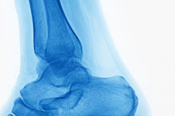
X-rays can rule out a fracture, but not assess for soft tissue injuries. Ultrasound can be used to assess the ankle ligaments. In one recent study, ultrasound had a sensitivity and specificity of 100% in distinguishing normal ATFL from a torn or sprained ligament. Clinical tests (the anterior drawer) had a sensitivity and specificity of 81% and 80% (Hosseinian et al, 2022).
In a systematic review and meta-analysis, sensitivity and specificity was 99% and 92% respectively (Seok et al, 2020). In this study, the authors recommend ultrasound as a first-line diagnostic tool to diagnose anterior tibiofibular ligament (ATFL) and calcaneofibular ligament (CFL) injuries.
Without proper treatment and rehabilitation, severe sprains can weaken your ankle and makes it more likely that you will injure it again. Repeated ankle sprains can lead to long-term problems, including chronic ankle pain, arthritis, and ongoing instability. Ultrasound can help guide management.
In a recent study by Delzell et al., the authors found that patients that presented with an acute ankle injury and had a diagnostic ultrasound the diagnosis changed in 64.3%. In this study, the treatment was affected after the ultrasound in 42.8% of cases.
In Delzell et al
The authors found that ultrasound helped avoid unnecessary surgery or injections, and changed conservative management
In another study, Sanjay et al. found that ultrasound helped guide whether to use bracing in acute injuries. This resulted in a higher rate of complete healing compared to other studies.
In mild ankle sprains, rehabilitation can begin within a few days of the injury when the swelling improves and focus on restoring mobility, strength and balance. Ultrasound can help guide management, and in severe ankle sprain athletes may want to consider taping the ankle or using a brace can help prevent re-injury.
Contact us for an ultrasound assisted diagnosis and informed treatment plan.
Boston Sports & Biologics
20 Walnut St., Suite #14
Wellesley Hills MA 02481
(781) 591-7855
References:
Learn how ultrasound-guided percutaneous fasciotomy may revolutionize treatment for chronic exertional compartment syndrome with faster recovery and smaller incisions.
Read MoreResearchers used artificial intelligence and natural language processing to identify Shoulder Injury Related to Vaccine Administration (SIRVA) cases from over 3.7 million vaccinations. Learn what the study found, how
Read More