Calf Strain with Hematoma: A Real Case of Gastrocnemius Injury and What the Evidence Says About Aspiration
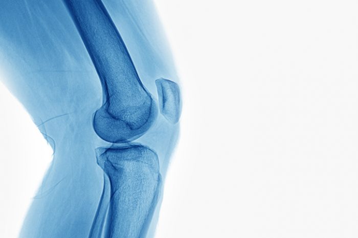
A calf strain, particularly of the medial head of the gastrocnemius, is a common soft tissue injury—especially in middle-aged athletes. Often called "tennis leg," it typically results from a sudden eccentric contraction, like pushing off for a sprint or jump.
What are common symptoms of a Gastrocnemius Strain (a.k.a. “Tennis Leg”)?
Common Symptoms:
Sudden sharp pain or “popping” sensation
Swelling and bruising
Difficulty weight-bearing
Tenderness over the medial calf
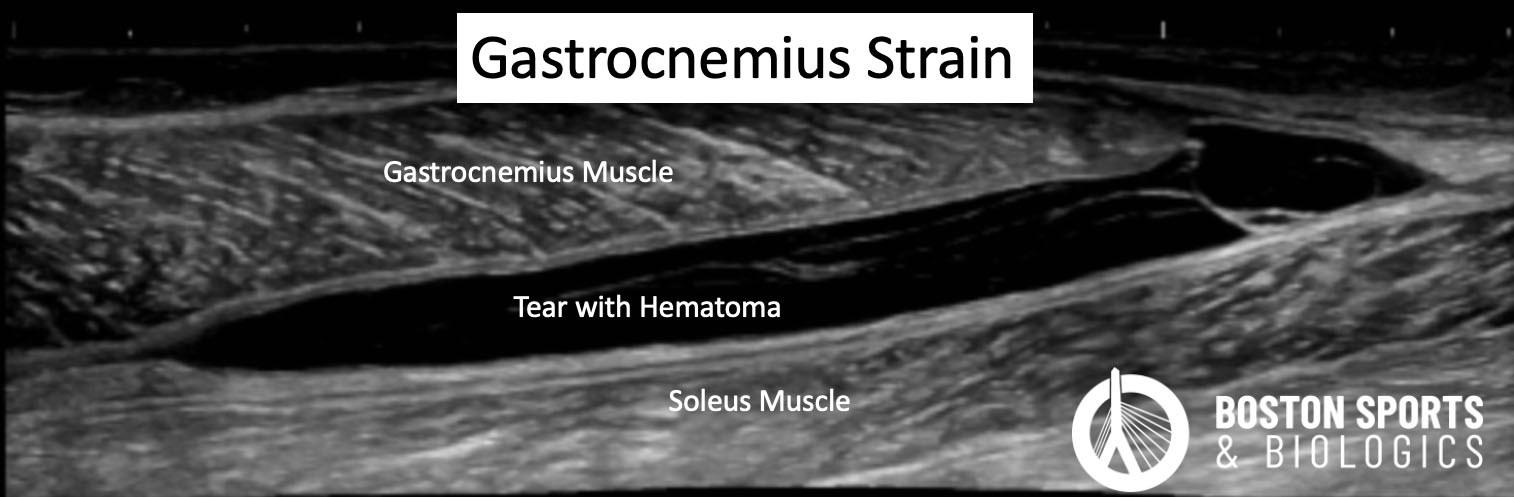
When bleeding occurs within the muscle belly, it can lead to a gastrocnemius hematoma—a localized collection of blood that can prolong recovery and increase discomfort.
What is the Role for Diagnostic Ultrasound in Calf Strains?
Most calf strains can be diagnosed through clinical history and exam. However, ultrasound or MRI may be used to:
Confirm the location and grade of the tear
Identify hematomas or fluid collections
Rule out other injuries (e.g., deep vein thrombosis—an important differential diagnosis!)
Ultrasound
is a valuable tool for diagnosing gastrocnemius strain due to its
non-invasive nature, cost-effectiveness, and ability to provide
real-time imaging. Several studies have demonstrated its efficacy in
identifying and classifying muscle injuries, including those of the
gastrocnemius.
Ultrasound Classification of Gastrocnemius Injuries
High-resolution ultrasound can detect both partial and complete tears of the gastrocnemius muscle, as well as associated hematomas and fluid collections[Yoon et al, 2021].
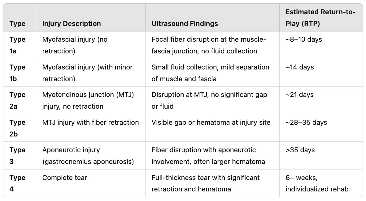
For instance, Pedret et al. proposed a classification system for medial gastrocnemius injuries based on sonographic findings, which correlated significantly with return-to-play and return-to-work times [Pedret et al, 2020].
This classification includes various types of injuries, such as myoaponeurotic injuries and injuries involving the gastrocnemius aponeurosis.
What the Evidence Says About Calf Strain Management
Conservative Treatment
Most Grade I and II calf strains respond well to nonoperative treatment:
RICE protocol in the acute phase
Progressive rehabilitation including stretching, eccentric strengthening, and mobility work
Compression therapy may help resolve hematoma faster
Crutches or heel lifts may be needed initially for pain relief
Evidence shows that early, structured rehab can accelerate return to play and minimize the risk of re-injury [Bayer et al, 2018].
Imaging-Guided Aspiration
In cases with significant hematoma, ultrasound-guided aspiration of musculoskeletal hematomas has been shown to be safe and effective, providing symptomatic relief without significant complications [Yoon et al, 2021; Szopinski & Smigielski, 2012].
Ultrasound-guided aspiration can help:
Decompress the muscle
Reduce pain and pressure
Improve mobility and speed up healing
While not routine, aspiration is supported by small studies and case reports showing earlier functional recovery and reduced fibrosis risk.
The literature indicates that early intervention, ideally within 3-5 days of hematoma formation, may improve outcomes and prevent complications such as compartment syndrome [De la Corte-Rodriguez, 2014; Mithöfer et al, 2002].
It is important to note that while aspiration can provide symptomatic relief, it may not always be successful, and there is a risk of hematoma recurrence [Szopinski & Smigielski, 2012].
Bayer ML, Hoegberget-Kalisz M, Jensen MH, Olesen JL, Svensson RB, Couppé C, Boesen M, Nybing JD, Kurt EY, Magnusson SP, Kjaer M. Role of tissue perfusion, muscle strength recovery, and pain in rehabilitation after acute muscle strain injury: Arandomized controlled trial comparing early and delayed rehabilitation. Scand J Med Sci Sports. 2018 Dec;28(12):2579-2591.
Cicvarić T, Sustić A, Miletić D, Veselko M, Mozetic V, Spanjol J. Endoscopic evacuation of a hematoma resulting from strain injury of the medial head of the gastrocnemius muscle. Arthroscopy. 2006 Aug;22(8):912.e1-3.
De la Corte-Rodriguez H, Rodriguez-Merchan EC. Treatment of muscle haematomas in haemophiliacs with special emphasis on percutaneous drainage. Blood Coagul Fibrinolysis. 2014 Dec;25(8):787-94.
Mithöfer K, Lhowe DW, Altman GT. Delayed presentation of acute compartment syndrome after contusion of the thigh. J Orthop Trauma. 2002 Jul;16(6):436-8.
Pedret C, Balius R, Blasi M, Dávila F, Aramendi JF, Masci L, de la Fuente J. Ultrasound classification of medial gastrocnemious injuries. Scand J Med Sci Sports. 2020 Dec;30(12):2456-2465.
Szopinski KT, Smigielski R. Safety of sonographically guided aspiration of intramuscular, bursal, articular and subcutaneous hematomas. Eur J Radiol. 2012 Jul;81(7):1581-3.
Yoon ES, Lin B, Miller TT. Ultrasound of Musculoskeletal Hematomas: Relationship of Sonographic Appearance to Age and Ease of Aspiration. AJR Am J Roentgenol. 2021 Jan;216(1):125-130.
Learn how ultrasound-guided percutaneous fasciotomy may revolutionize treatment for chronic exertional compartment syndrome with faster recovery and smaller incisions.
Researchers used artificial intelligence and natural language processing to identify Shoulder Injury Related to Vaccine Administration (SIRVA) cases from over 3.7 million vaccinations. Learn what the study found, how