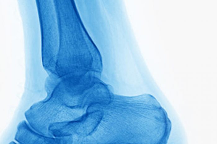

Fat pad atrophy affects nearly 30% of individuals over 60 [Hannan et al, 2013], making simple activities like walking a painful ordeal.
In a groundbreaking study titled “Volumetric Analysis in Autologous Fat Grafting to the Foot” by Ruane et al. delves into the science behind a forefoot fat grafting procedure, shedding light on its efficacy in alleviating pain and restoring function in individuals suffering from foot fat pad atrophy.
This study is the first to use MRI analysis to measure the effectiveness of fat grafting in restoring foot pad volume and relieving pain.
The natural cushioning under our feet, known as the plantar fat pad, plays a crucial role in absorbing shock and reducing pressure on our bones. However, due to aging, repeated impact, steroid use, or certain medical conditions, this protective layer thins over time, leading to direct pressure on the bones of the foot. The result? Pain, calluses, ulcers, and even changes in gait that can impact overall mobility and quality of life.
Traditional treatments such as padding and synthetic fillers provide temporary relief, but they do not restore lost volume effectively. This is where autologous fat grafting comes in as a promising, minimally invasive alternative.
Ruane et al. conducted a prospective study using MRI technology to analyze 3D volume changes in the foot before and after autologous fat grafting. Seventeen participants (six men and eleven women) suffering from fat pad atrophy underwent the procedure, receiving an average of 5.8 cc of fat injections per foot.
The key findings of the study include:
While previous studies using ultrasound suggested that fat grafted under the metatarsal heads disappears within 2–6 months, MRI analysis in this study tells a different story. Instead of vanishing, the fat redistributes, providing long-term support around the metatarsal heads. This could explain why pain relief extends up to two years despite measurable reductions in direct tissue thickness.
Additionally, fat grafting offers biological benefits beyond volume restoration. The grafted fat contains stem cells, which may contribute to tissue regeneration, improving the biomechanical properties of the plantar pad. The authors are currently investigating the stem cell characteristics of the fat used in these foot fat grafting cases and aim to correlate it to clinical findings in the near future [Ruane et al, 2019].
While this study provides compelling evidence of the benefits of autologous fat grafting, further research is needed to assess long-term fat retention. Exploring the regenerative potential of stem cells within grafted fat could unlock new frontiers in foot care.
As advancements in regenerative medicine continue, autologous fat grafting is poised to become a mainstream treatment for foot pain, offering a natural and effective solution for millions suffering from fat pad atrophy [Ruane et al, 2019].
The study by Ruane et al. reaffirms what many in the medical community have suspected—autologous fat grafting is a game-changing technique for foot pain management. With proven increases in foot pad volume, pain reduction, and functional improvement [Ruane et al, 2019]..
Have you experienced foot pain or undergone fat grafting? Learn more at:
References:
Meniscus tears are one of the most common causes of knee pain in active adults and aging athletes alike. If you’ve been told you have a “degenerative meniscus tear,” you may have also heard that surgery isn’t always the
Read MoreFor patients with chronic adductor longus tendinopathy, a newer option is emerging: ultrasound-guided tenotomy using the Tenex system. Recent clinical evidence suggests this minimally invasive approach may effectively
Read More