Email: info@BSBortho.com
Trigger finger, also known as stenosing tenosynovitis, is a common condition that causes pain, stiffness, catching, or locking of a finger or thumb. Trigger finger (stenosing tenosynovitis) occurs due to a size discrepancy between the flexor tendon and the flexor tendon sheath in which it resides, the tendon becomes larger or wider while the sheath becomes smaller or narrower, preventing smooth gliding as the finger bends and straightens (Currie et al, 2022).
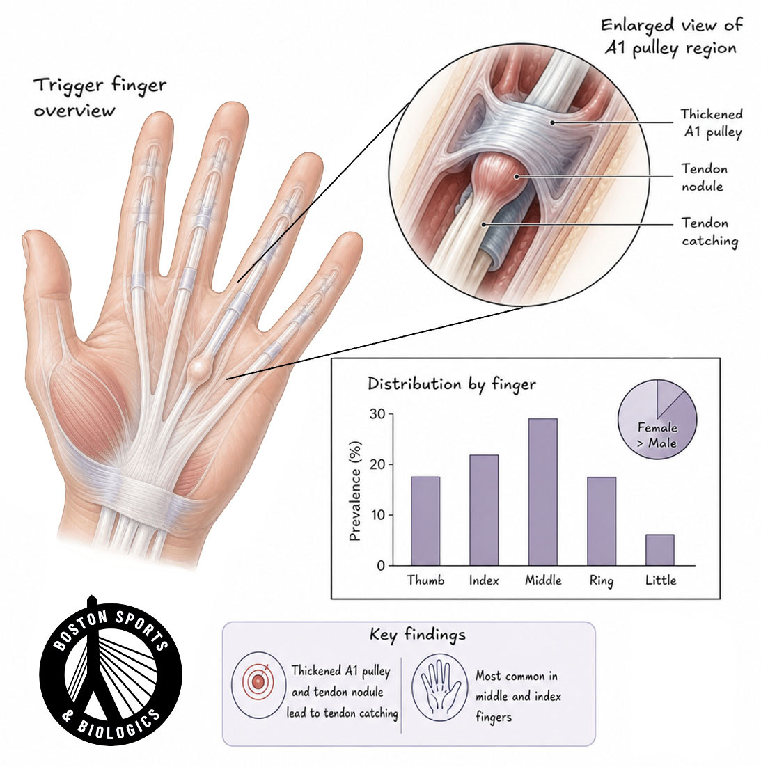
Trigger finger typically presents initially with subtle pain along the A1 pulley distribution, crepitus with range of motion, or a palpable tender nodule along the tendon. More advanced disease presents with inability to fully flex or extend
the finger, locking of the finger in flexion, and painful popping with flexion or extension.
Trigger finger can affect any finger. The condition affects approximately 2% of the general population and up to 20% of people with diabetes (Currie et al, 2022).
The flexor tendons are cord-like structures that connect the forearm muscles to the fingers and thumb, allowing the hand to grip and grasp objects. These tendons travel through a series of fibrous tunnels called pulleys that keep the tendon close to the bone during motion.
The A1 pulley, located near the base of the finger in the palm, is the most common site involved in trigger finger.
The friction caused by flexor tendons passing through the stenotic A1 pulley can change the tendon fibers, forming an intratendinous lump (nodule). When the tendon becomes thickened or the pulley becomes constricted, the tendon can no longer glide smoothly through the pulley. This creates the characteristic catching, clicking, and locking symptoms associated with trigger finger.
The exact cause is not always known, but several factors increase the risk of developing trigger finger (Fiorini et al, 2018). Risk factors include occupations or activities requiring repetitive gripping, metabolic disorders (e.g., diabetes), obesity, age between 40 and 60 years, female sex, and inflammatory arthritis.
Diabetes mellitus: Trigger finger affects up to 20% of people with diabetes, with cross-sectional studies reporting prevalences of 4%–55%. Higher HbA1c levels are independently associated with increased risk in both type 1 and type 2 diabetes, suggesting hyperglycemia directly contributes to development (Currie et al, 2022; Rydberg et al, 2022).
Rheumatoid arthritis: Identified as a significant risk factor in a large population-based cohort study, along with female sex and age 50–59 years (Shen et al., 2019).
Carpal tunnel syndrome: A prospective study found that 43% of patients with trigger finger had clinical signs of carpal tunnel syndrome, and 61% of patients with either condition had both on examination (Patel et a., 2022).
Dupuytren's disease: Listed as part of the differential diagnosis and as an associated condition, with trigger finger release surgery itself identified as a risk factor for subsequent Dupuytren's disease development (Currie et al, 2022; Novo et al, 2026).
Other associated conditions: Gout, De Quervain's disease, amyloidosis, mucopolysaccharidosis, hypothyroidism, and congestive heart failure have all been linked to trigger finger as a consequence of connective tissue metabolism changes (Fiorini et al, 2018).
Prior hand injury: Distal radius fractures treated with ORIF were associated with subsequent trigger finger development, with diabetes as a significant additional risk factor (Yeh et al, 2020).
Repetitive hand use: Activity modification (minimizing repetitive gripping or pinching) is a first-line nonoperative treatment, underscoring the role of repetitive use in symptom exacerbation.
Trigger finger is more common in women and typically occurs between the ages of 40 and 70, and is four to six times more frequent in women than men (Fiorini et al, 2018).
Symptoms often develop gradually. Trigger finger typically presents with:
Pain at the base of the finger: Subtle pain along the A1 pulley distribution in the palm at the metacarpal head (Currie et al, 2022).
Tenderness in the palm and palpable nodule: A palpable tender nodule along the tendon or thickening is a characteristic finding (Currie et al, 2022).
Clicking, popping, and catching: The clinical manifestation may vary from occasional snapping to a finger locked in flexion, caused by the friction of flexor tendons passing through the A1 (Fiorini et al, 2018).
Stiffness, especially in the morning: Symptoms may start as pain in the palm or proximal interphalangeal joint without triggering, then increase in severity, leading to stiffness of the affected PIP joint (Fiorini et al, 2018).
Locking in a bent position: More advanced disease presents with locking of the finger in flexion and painful popping with flexion or extension, sometimes requiring manual unlocking with the opposite hand (Currie et al, 2022).
Difficulty gripping: Trigger finger is classified as an occupational overuse syndrome occurring among people whose jobs require repetitive gripping.
Symptoms frequently worsen with repetitive hand use.
Trigger finger is primarily a clinical diagnosis based on a patient's symptoms and physical examination.
Physical Examination Findings
The examination elements listed in the text are well supported:
Finger motion and triggering: Physical examination demonstrates locking and catching at the affected metacarpophalangeal joint as the patient flexes and extends the fingers (Pujalte et al, 2024).
Tenderness over the A1 pulley: The physician should assess for pain or tenderness at the finger's base, a tender nodule, or palmar fascial thickening. Clinical examination typically demonstrates tenderness and nodularity of the flexor tendon sheath near the palmar A1 pulley and associated motion-based catching or locking (Pujalte et al, 2024; Hall & Compton, 2025).
Finger locking: More advanced disease presents with inability to fully flex or extend the finger, locking of the finger in flexion, and painful popping with flexion or extension (Currie et al, 2022).
Associated hand conditions: The differential diagnosis includes infection, extensor tendinitis, arthritis, Dupuytren contracture, and carpal tunnel syndrome, all of which should be assessed during evaluation (Currie et al, 2022).
Diagnostic ultrasound provides an excellent method for evaluating trigger finger and may demonstrate:
Ultrasound also helps identify other causes of finger pain and allows precise guidance for therapeutic procedures.
Corticosteroid injection has long been considered a first-line intervention for trigger finger.
Ultrasound guidance allows precise placement of medication around the flexor tendon sheath while avoiding injury to nearby structures.
Potential benefits include:
Success rates are generally highest in patients with shorter symptom duration and less severe locking.
However, recurrence is common, particularly in patients with diabetes or longstanding symptoms.
For patients with persistent symptoms despite conservative treatment, release of the A1 pulley may be recommended.
Traditionally, trigger finger release has been performed through an open surgical incision in the palm. While highly successful, open surgery requires an incision, postoperative wound care, and a recovery period before returning to unrestricted activities.
Ultrasound-guided A1 pulley release is a minimally invasive alternative to open surgery.
Ultrasound-guided trigger finger release is a minimally invasive procedure performed through a tiny skin opening using real-time ultrasound visualization.
Using ultrasound guidance, the physician can:
Ultrasound allows visualization of the A1 pulley, identification of flexor tendons, avoidance of nerves and blood vessels, and confirmation of complete release. Because the procedure is performed under continuous imaging guidance, surrounding structures can be protected throughout the procedure.
Potential advantages compared with traditional open surgery include:
Many patients are able to begin using their hand almost immediately following the procedure.
You may be a candidate if you:
A consultation and ultrasound evaluation can help determine the most appropriate treatment approach.