Ultrasound-guided foreign body removal is a nonsurgical highly effective technique and should be considered as a first-line treatment procedure. Various types of foreign bodies can be diagnosed and removed with ultrasound, including medical devices such as contraceptive implants, wooden splinter, metallic objects, and glass objects, and they may be medical devices such as contraceptive implants. Frequently, physical examination is not sensitive enough to detect a foreign body, and ultrasound is the imaging technique of choice and the effectiveness of ultrasound-guided percutaneous removal can be near 100%.
A soft tissue foreign body is an outside object that gets embedded in the tissue under the skin, and can be a splinter, rock or piece of metal or shard of glass. The location of the foreign body can vary, and the only symptoms may be pain, local swelling, or signs of an inflammatory response.
Soft-tissue foreign bodies can result from accidents or medical procedures, such as a contraceptive implants.
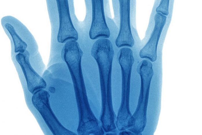
Foreign bodies can be difficult to feel or detect on physical examination. Often imaging is used to localize the object, but not all foreign bodies will be seen on radiographs. Traditionally, patients suspected of having a foreign body undergo imaging. Metallic objects like nails and pins can usually be detected on x-rays. Depending on size and location, even some normally visible objects may be hard to visualize. Other common objects, such as wood/splinters or glass are difficult to see
If x-rays fail to identify a retained soft tissue foreign body and one is suspected, ultrasound is the next imaging modality of choice. Ultrasound has a high specificity (92%) and sensitivity to identify foreign bodies (Davis et al. 2015).
Foreign bodies need to be removed, and failure to remove foreign bodies can give rise to acute pain and/or functional impairment or late complications, such as inflammation or infection. The surgical removal of foreign bodies is invasive, costly and technically challenging requiring a relatively large incision.
Ultrasound can be used to help guide removal of foreign bodies. Percutaneous ultrasound-guided foreign body removal was first described in 1990 (Shiels et al, 1990). Ultrasound can help accurately localize a foreign body, and remove the foreign body in a minimally invasive way that decreases the amount of bleeding and helps avoid injuring adjacent structures.
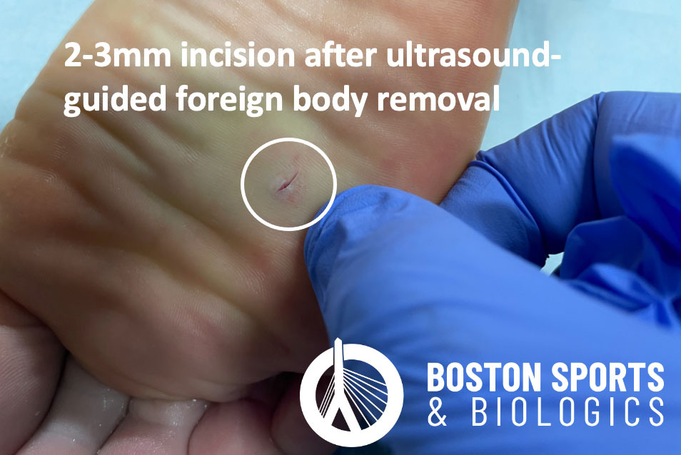
Ultrasound-guidance allows the object to be removed through a small incision, which minimizes the risk of infection and the decreases the aesthetic impact.
This patient had a splinter removed 1-year before presenting to our clinic. Her primary care physician had removed the splinter, but she still felt like something was in her foot. She had x-rays and n MRI that did not show the retained splinter, but the splinter can easily be seen on ultrasound.
We then decided to use ultrasound to help remove the retained splinter. The splinter was removed using local anesthetic injection, a small incision in the skin and the small retained wood splinter was removed through the incision. The incision was too small to need stitches and was covered with adhesive strips.

To schedule a consultation and learn if you are a candidate, call to schedule:
For patients with chronic adductor longus tendinopathy, a newer option is emerging: ultrasound-guided tenotomy using the Tenex system. Recent clinical evidence suggests this minimally invasive approach may effectively
Read MoreAdductor longus selective tenotomy is a modern surgical treatment for chronic groin pain that offers faster recovery and better outcomes than traditional full release surgery. The adductor longus, an inner thigh
Read More