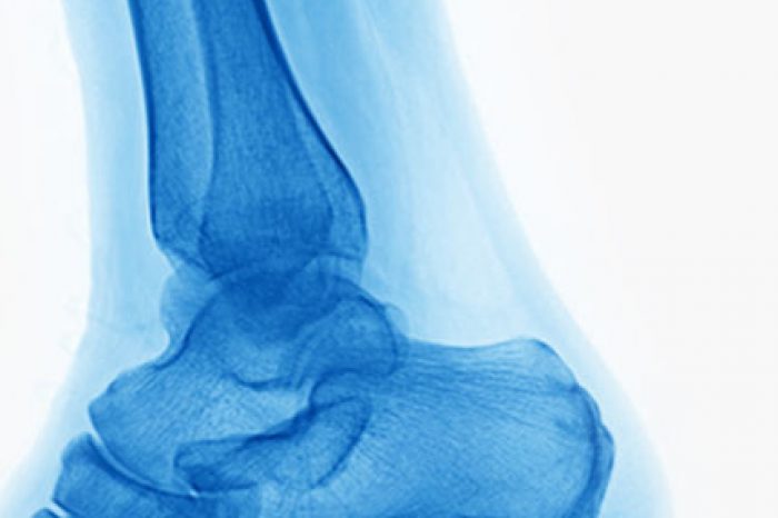
Plantar fat pad atrophy is a common but under diagnosed cause of persistent ball-of-foot pain. This condition happens when the natural fat pad under the metatarsal heads (the ball of your foot) or the heel breaks down, leading to a painful sensation that feels like you are “walking on bone.”
Traditionally, plantar fat pad atrophy has been a diagnosis of exclusion, meaning doctors ruled out other problems first. But today, advances in musculoskeletal ultrasound make it possible to directly measure and monitor the fat pad, helping patients get a quicker, more accurate diagnosis.
The fat pad under your forefoot acts like a natural shock absorber, protecting bones and joints from repetitive impact. When it deteriorates due to aging, prior surgery, steroid injections, or mechanical overload, the cushioning effect weakens.
The result? More pressure on bone and soft tissue, leading to pain, callus formation, and even ulcers in high-risk groups like people with diabetes.
Unlike X-rays, which only show bone, ultrasound gives a real-time picture of soft tissues—including the plantar fat pad.
Key benefits include:
Direct measurement of thickness: Ultrasound can determine if the fat pad is below healthy thresholds. Research suggests that a plantar fat pad thickness in the forefoot of less <0.7 cm is a red flag for atrophy.
Detection of degeneration: Ultrasound shows changes in fat quality, such as thinning or fibrosis (scarring), that reduce its cushioning ability.
Dynamic assessment: Doctors can evaluate how the fat pad behaves dynamically but putting pressure during the study. This dynamic examination can mimic weight bearing and real walking conditions.
Comparisons to the other foot: Ultrasound allows side-by-side measurement with your unaffected foot for context.
A recent study from the University of Seville found that patients with predislocation syndrome (a painful instability of the toe joint) had much thinner plantar fat pads (0.57 cm) compared to healthy controls (0.94 cm), confirming ultrasound’s role in linking fat pad loss to forefoot pain.
Consider asking your doctor about an ultrasound evaluation if you have:
Persistent pain in the ball of the foot not explained by arthritis, bunions, or neuromas
A history of steroid injections or foot surgery
A sensation of “walking on pebbles” or lack of cushioning
Diabetes or rheumatoid arthritis, conditions that increase risk of fat pad thinning
Painful calluses or early ulceration despite using orthotics or padding
The exam is simple, quick, and painless. A small probe glides across the ball of your foot with ultrasound gel, sending sound waves that create images of the fat pad in real time.
Your provider may measure:
Plantar fat pad thickness beneath each metatarsal head
Plantar plate thickness, a ligament-like structure that stabilizes your toes, which can also be affected
Fat quality, noting whether the tissue is uniform and healthy or fibrotic
Once diagnosed, ultrasound can:
Track progression of atrophy over time
Guide injections or fat grafting procedures with precision
Measure success after treatment such as autologous fat grafting, dermal grafts, or fillers
For example, a prospective clinical trial at the University of Pittsburgh used ultrasound to measure fat pad thickness before and after autologous fat grafting. Patients reported less pain and better function after the procedure, even though ultrasound showed gradual thinning of the graft over 6–12 months.
While MRI can also visualize the fat pad, ultrasound is faster, more affordable, and available right in the office. For most patients, it is the first-line imaging tool for diagnosing plantar fat pad atrophy.
Yes. Ultrasound uses sound waves, not radiation, making it completely safe and repeatable.
A typical exam lasts 10–15 minutes.
No. The probe only touches the skin, though you may feel mild pressure.
Studies suggest >0.8–0.9 cm is typical. Anything <0.7 cm indicates atrophy and higher risk for pain or complications.
Yes—advanced ultrasound techniques like elastography can assess tissue stiffness and elasticity, giving more detail about cushioning ability.
Yes. Orthotics remain the mainstay of treatment, but ultrasound helps your doctor decide if you may also benefit from advanced options like fat grafting.
Left untreated, plantar fat pad atrophy can worsen, leading to more pain, limited mobility, and even ulcers. With ultrasound, clinicians can identify the condition early, start protective measures like custom orthotics, and discuss restorative treatments if needed.
Ultrasound is transforming the way doctors diagnose plantar fat pad atrophy. By directly measuring fat thickness, tracking degeneration, and guiding treatment, this safe, non-invasive tool helps patients move from “mystery pain” to answers—and, most importantly, relief.
If you’re struggling with persistent forefoot pain, ask your foot and ankle specialist whether an ultrasound evaluation of your plantar fat pad could provide the clarity you need.
(781) 591-7855
20 Walnut St
Suite 14
Wellesley MA 02481
Gusenoff JA, et al. Autologous Fat Grafting for Pedal Fat Pad Atrophy: A Prospective Randomized Clinical Trial. Plast Reconstr Surg. 2016;138(5):1099–1107. PubMed
Rocchio TM. Augmentation of Atrophic Plantar Soft Tissue with an Acellular Dermal Allograft: A Series Review. Clin Podiatr Med Surg. 2009;26(4):545–557. PubMed
Rayo Pérez AM, et al. Ultrasound Relationship of Plantar Fat and Predislocation Syndrome. Diseases. 2025;13(5):128. Full Text
Is stem cell therapy better than cortisone for knee arthritis? Learn the differences, risks, benefits, and long-term results of cortisone vs stem cell knee injections.
Read More