Email: info@BSBortho.com

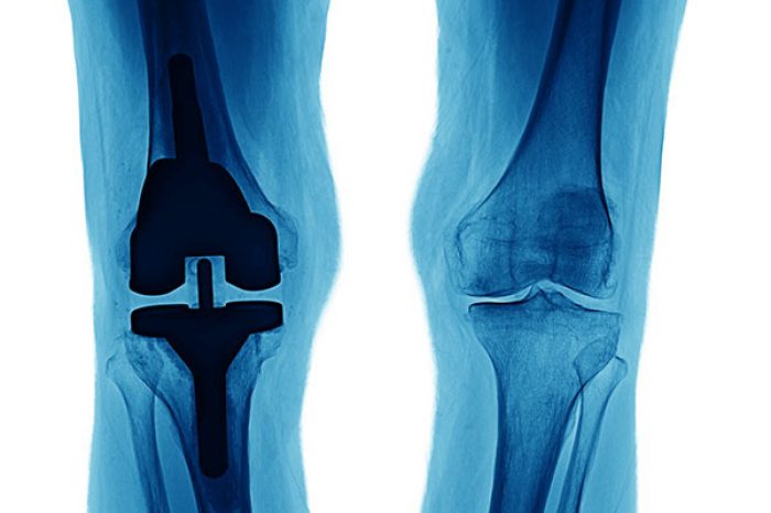
When people think about knee osteoarthritis (OA), they often think about cartilage: the smooth tissue covering the ends of the bones becomes damaged or thinned over time. But cartilage is only one part of the story.
Increasing evidence suggests that the bone immediately beneath the cartilage, called subchondral bone, can also play an important role in osteoarthritis and knee pain. One finding that physicians may see on a knee MRI is a bone marrow lesion (BML).
Understanding bone marrow lesions may help explain an important question many patients with knee arthritis have: Why does my knee hurt so much when I walk or stand?
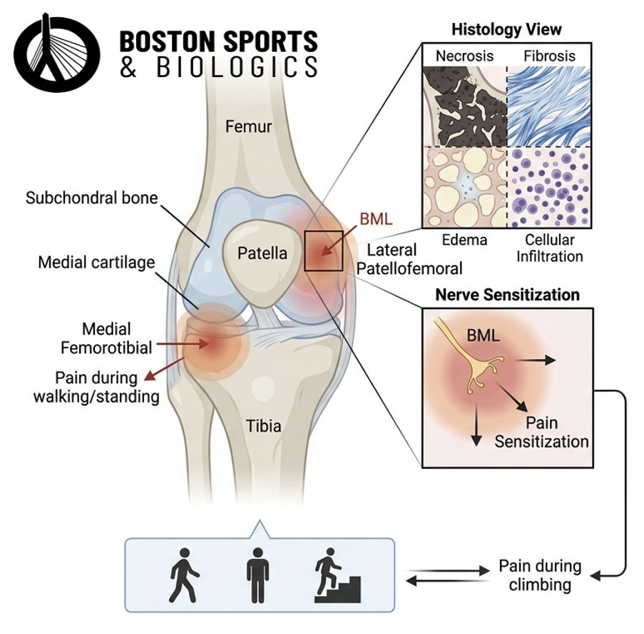
A bone marrow lesion is an area of abnormal signal within the bone that can be seen on an MRI. BMLs commonly occur in the subchondral bone. The subchondral bone sits directly beneath the cartilage and provides a shock-absorbing function while supplying nutrients to deeper cartilage layers through its micropores. Distinctive OA changes here include sclerosis, trabecular remodeling, osteophytes, bone cysts, and bone marrow lesions. Some evidence suggests subchondral bone changes may even precede cartilage degeneration (Wu et al, 2022).
Although sometimes informally described as "bone marrow edema," a BML is more complicated than simply swelling or fluid in the bone. Histologic studies showed edema is only a minor constituent. Studies examining these areas under a microscope have identified changes that can include abnormal bone remodeling, fibrosis, vascular changes, cellular infiltration, and areas of bone marrow damage (Muratovic et al, 2018).
In other words, a BML can be thought of as an MRI sign that the bone underneath an arthritic joint is undergoing an abnormal response to stress and injury.
For years, knee arthritis treatment focused heavily on cartilage loss. We now understand that osteoarthritis involves the entire joint, including cartilage, bone, meniscus, synovium, ligaments, and other tissues (Goldring & Goldring, 2016).
Subchondral bone is particularly interesting because, unlike cartilage, bone contains nerves capable of transmitting pain (Aso et al, 2019).
Bone marrow lesions are more common in people with knee OA than in people without arthritis, and larger BMLs have been associated with greater knee pain. Studies have also found that increasing BML volume over time can be associated with increasing pain (Klement & Sharkey, 2019; Xu et al, 2012).
This does not mean that every BML causes pain. Some people can have bone marrow lesions without knee symptoms. Rather, the location, size, underlying biomechanics, and other abnormalities in the knee all appear to matter (Tomura et al, 2025).
One of the most interesting studies of this issue examined data from the Osteoarthritis Initiative, a large research project studying knee osteoarthritis.
Aso and colleagues analyzed 1,412 knees and followed 582 knees with bone marrow lesions over 24 months (Aso et al, 2021). They examined BMLs in different regions of the knee and compared them with pain during weight-bearing activities.
The researchers separated pain into two categories.
Weight-bearing pain included pain while:
Non-weight-bearing pain included pain while sitting, lying down, or in bed. This distinction is important because pain that occurs primarily when the knee is loaded may have a different source than pain occurring at rest.
Bone marrow lesions in the medial femorotibial compartment, the inner portion of the knee where the femur meets the tibia, were associated with greater weight-bearing pain.
More specifically, larger lesions in this area were associated with more pain during walking and standing. BMLs in the anterior knee (the lateral patellofemoral region) were associated with pain while climbing stairs.
Even more interestingly, when medial femorotibial BMLs increased or decreased over 24 months, weight-bearing pain tended to increase or decrease as well. These findings remained significant even after the researchers accounted for several other findings commonly seen with knee OA.
This supports the idea that, for some patients, the painful structure may not simply be the cartilage, it may also be the bone underneath it.
Mechanical overload appears to be one important contributor (Beckwée et al, 2015).
When the knee does not distribute forces normally, certain areas of subchondral bone may experience greater stress. For example, a bow-legged or varus knee typically places greater load through the medial compartment.
BMLs have also been associated with factors including:
Meniscus and cartilage normally help distribute and absorb forces across the knee. When these structures become damaged, greater forces may be transferred to the underlying bone. This creates a more complete way of thinking about knee OA: cartilage damage, meniscal dysfunction, abnormal joint mechanics, and subchondral bone changes can interact with one another (Aso et al, 2021).
Potentially, but treatment should be individualized.
The presence of a BML does not automatically mean that it needs a specific procedure. It may instead provide another piece of information about where pain is coming from and how forces are being distributed through the knee (Walsh et al, 2023).
For example, treatment may include exercise and rehabilitation, weight management when appropriate, activity modification, bracing, and strategies designed to improve joint mechanics or reduce excessive loading of an affected compartment.
There is evidence that mechanical unloading can influence some BMLs. Patellofemoral bracing reduced BML volume in patients with painful patellofemoral OA (Callaghan et al, 2015). However, other approaches, such as lateral wedge insoles for medial knee OA, have not consistently reduced BMLs or pain (Aso et al, 2021).
Importantly, knee OA has multiple potential sources of pain. Treating the subchondral bone would therefore be expected to be most relevant when the BML and underlying bone appear to be important contributors to a patient's symptoms.
Knee osteoarthritis is more than simply “wearing out the cartilage.”
The bone underneath the cartilage matters, and bone marrow lesions seen on MRI may be an important source or marker of pain in certain patients. Research suggests that BMLs in weight-bearing portions of the knee are particularly associated with pain during activities such as standing and walking. Changes in these lesions over time may also parallel changes in pain.
This is one reason why evaluating knee arthritis should involve more than simply looking at the amount of “joint space narrowing” on an X-ray. Understanding which structures are abnormal, where they are located, and how the knee is being loaded can help provide a more complete picture of why an individual patient's knee hurts.
The terms are sometimes used interchangeably, but bone marrow lesion is generally the more accurate term. These MRI abnormalities can represent several tissue changes rather than simply fluid or swelling. Histologic studies have identified fibrosis, abnormal trabecular bone, vascular changes, cellular infiltration, and other abnormalities within BMLs.
They can be associated with pain, particularly when located in weight-bearing portions of an arthritic knee. However, not every bone marrow lesion is symptomatic, and bone marrow lesions can occur in people without knee pain.
There can be several reasons. One possibility is abnormal stress within the subchondral bone. Research has found an association between medial femorotibial bone marrow lesions and pain during walking and standing.
Bone marrow lesions can change over time. In this Aso study, both increases and decreases in medial femorotibial bone marrow lesion severity over 24 months were associated with corresponding changes in weight-bearing pain.
Not necessarily. Many patients can be evaluated using their history, physical examination, and X-rays. MRI can provide additional information about cartilage, menisci, ligaments, subchondral bone, and bone marrow lesions when these structures may affect diagnosis or treatment.
No. A bone marrow lesion is an imaging finding, not an automatic indication for surgery. Its importance depends on its location, severity, symptoms, alignment, and the other abnormalities present in the knee.
Possibly. Mechanical loading appears to influence bone marrow lesion development and progression, and some unloading strategies have reduced bone marrow lesion size in research studies. However, the amount and type of unloading required, and which patients benefit, remain uncertain.

(781) 591-7855
20 Walnut St
Suite 14
Wellesley MA 02481
How safe are PRP injections for knee arthritis? A recent study found that PRP has similar side effects to hyaluronic acid and steroid injections, with temporary post-injection pain being the most common reaction.
Read MoreKnee arthritis isn't just about cartilage. Bone marrow lesions in the bone beneath the cartilage may contribute to pain with walking, standing, and stairs. Learn what these MRI findings mean and why their location
Read More