Email: info@BSBortho.com
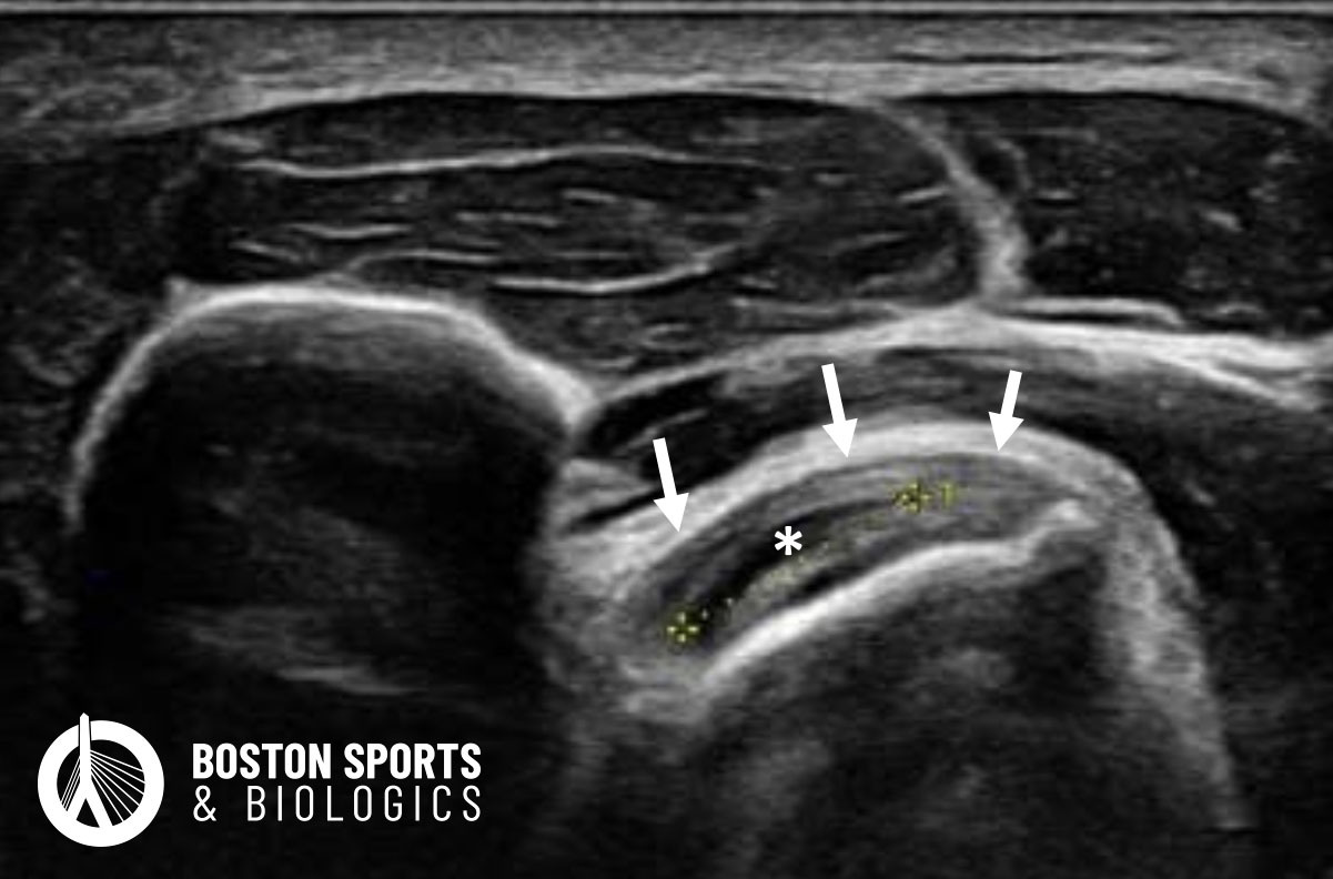
Distal biceps tendinopathy
refers to a condition affecting the the
biceps brachii tendon at the elbow. Biceps tendinopathy at the elbow is
primarily caused by a combination of mechanical, include overuse,
repetitive strain, or acute trauma, and anatomical factors.
Mechanical impingement: The
distal bicep tendon inserts into the radial tuberosity at the elbow,
and during forearm pronation there is increased pressure on the distal
biceps tendon [Rausch et al, 2020].
Anatomical factors can include a larger radial tuberosities and smaller radioulnar spaces predisposing the tendon to tendinopathy or tears [Boyle et al, 2022].
Overuse and repetitive strain are additional contributing factors. Chronic overuse can lead to microtrauma and degenerative changes within the tendon, overwhelming its reparative capacity and resulting in tendinopathy [Järvinen et al, 1997; Almekinders et al, 2003].
Disorganization of Collagen Fibers: The collagen fibers in the tendon become discontinuous and disorganized, losing their normal parallel alignment.
Increased Ground Substance: There is an increase in the amount of mucoid degeneration, indicative of extracellular matrix dysregulation.
Cellular Changes: Tenocytes, the resident cells in the tendon, often become enlarged (hypertrophic) and there is also an increase in cellularity, with a conspicuouspresence of fibroblasts and myofibroblasts.
Absence of Inflammatory Cells: Unlike tendinitis, which involves inflammation, tendinopathy is generally a non-inflammatory condition. Histopathological examination typically shows an absence of inflammatory cells.
Barker et. al. studied 6 patients who underwent an ultrasound-guided PRP injection, which showed to be a safe and effective treatment for recalcitrant cases of distal biceps tendinopathy.
Learn more about PRP injections here
Surgical Management
Surgery may be indicated if there is a failure of conservative management, a job requiring a high level of physical demands or a desire to return to high physical activity levels. In cases with a larger tear involving greater than 50% on MRI may also be predictive of a need for surgical intervention [Bauer et al, 2018]. Surgical options include:
Lobo Lda G, Fessell DP, Miller BS, Kelly A, Lee JY, Brandon C, Jacobson JA. The role of sonography in differentiating full versus partial distal biceps tendon tears: correlation with surgical findings. AJR Am J Roentgenol. 2013 Jan;200(1):158-62.
Lynch J, Yu CC, Chen C, Muh S. Magnetic resonance imaging versus ultrasound in diagnosis of distal biceps tendon avulsion. Orthop Traumatol Surg Res. 2019 Sep;105(5):861-866.
McShane JM, Nazarian LN, Harwood MI. Sonographically guided percutaneous needle tenotomy for treatment of common extensor tendinosis in the elbow. J Ultrasound Med. 2006 Oct;25(10):1281-9. doi: 10.7863/jum.2006.25.10.1281. PMID: 16998100.
(781) 591-7855
20 Walnut St
Suite 14
Wellesley MA 02481