Follow us on

THROWER'S ELBOW

What is Thrower’s Elbow?

  • Thrower's elbow refers to a constellation of injuries affecting the elbow joint, commonly seen in athletes who engage in repetitive overhead throwing activities, such as baseball pitchers, football quarterbacks, and javelin throwers.

  • The condition arises due to the significant valgus stress and repetitive microtrauma imposed on the elbow during the throwing motion.

    • During the late phases of throwing, the elbow is exposed to high stress and an extreme valgus load on the inside (medial) elbow [Powell et. al., 2021]. Over time, this leads to degenerative changes due to the microtrauma occurring.
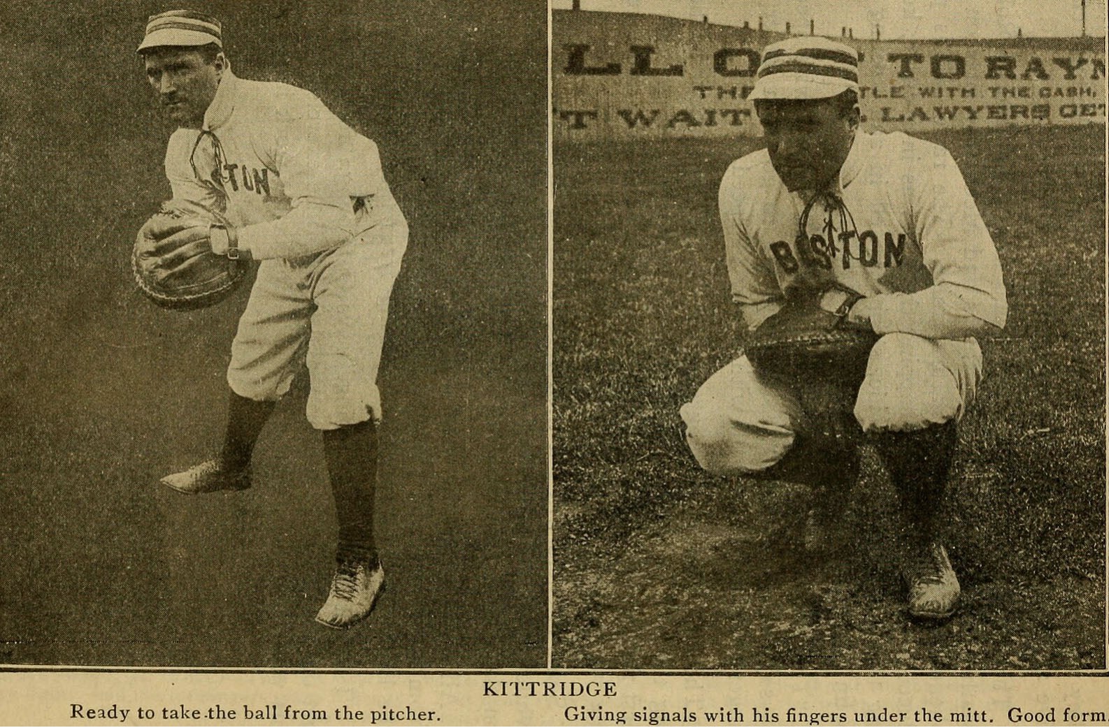
Contributing Library: The Library of Congress, Contributed by Wikimedia Commons (Public Domain)
  • The primary injuries associated with thrower's elbow include:
    • Ulnar Collateral Ligament (UCL) Injuries: The UCL is the main stabilizer against valgus stress. Repetitive throwing can lead to partial or complete tears of the UCL, resulting in valgus instability [Patel et al, 2014; Cain et al, 2003; Gehrman & Grandizio, 2022; Bruce & Andrews, 2014]. Learn more about UCL injuries HERE.
    • Flexor-Pronator Muscle Strain or Tendinitis: Overuse of the flexor-pronator muscle group can lead to tendinitis or muscle strain, contributing to medial elbow pain [Cain et al, 2003; Chen et al, 2001].
    • Medial Epicondyle Apophysitis or Avulsion: Particularly in younger athletes, repetitive stress can cause inflammation or avulsion of the medial epicondyle [Cain et al, 2003].
    • Valgus Extension Overload Syndrome: This condition involves the posterior compartment of the elbow, where repetitive valgus stress leads to osteophyte formation, chondromalacia, and loose bodies [Cain et al, 2003; Mller & Savoie, 1994].
    • Olecranon Stress Fractures: Repetitive stress can lead to stress fractures of the olecranon [Cain et al, 2003].
    • Ulnar Neuritis: The ulnar nerve can be affected by traction, friction, or compression due to the repetitive valgus stress [Cain et al, 2003; Chen et al, 2001].

How do you know if you have Thrower’s Elbow?

  • Thrower's elbow is a non-specific diagnosis and determining the underlying causes involves a detailed history, physical examination, and imaging.

    • History: A detailed history is crucial. This includes the duration and location of pain, the phase of the throwing motion during which pain occurs, and any associated symptoms such as decreased throwing velocity or control [Cain et al, 2003].

    • Physical Examination: The examination should include inspection, palpation, range of motion assessment, and specific tests. Key provocative maneuvers for UCL injury include the valgus stress test, the moving valgus stress test, and the milking maneuver. For flexor-pronator muscle strain or tendinitis, palpation over the medial epicondyle and resisted wrist flexion can
      elicit pain [Hariri & Safran, 2010; Ciccotti & Ciccotti, 2020; Hsu et al, 2012].

    • Imaging:

      • Ultrasound (US): Musculoskeletal US is valuable for dynamic assessment of tendons and ligaments. Stress US can detect UCL injuries by measuring medial joint space gapping under stress [Thomas et al, 2022; Hultman et al, 2021].

      • MRI and MR Arthrography: MRI is the gold standard for diagnosing UCL injuries, with MR arthrography providing higher accuracy for differentiating partial from complete tears. MRI is also useful for identifying other conditions such as medial epicondyle apophysitis, valgus extension overload syndrome, and olecranon stress fractures [Thomas et al, 2022].

      • Radiographs: Stress radiographs can measure medial joint space opening, which correlates with UCL injury severity [Thomas et al, 2022].

What are my treatment options?

  • Treatment options for thrower's elbow vary depending on the specific diagnosis and severity of the condition. Here is a detailed overview of the treatment strategies for the different conditions associated with thrower's elbow:
    • Ulnar Collateral Ligament (UCL) Injuries:
      • Nonoperative Management:Rest, physical therapy, and a graduated throwing program are first-linetreatments for low- to medium-grade partial UCL tears. Platelet-richplasma (PRP) injections may be considered [Carr et al, 2020; Marcaccio et al, 2025].
      • Surgical Management: Complete UCL tears or failure of conservative management may require UCL reconstruction, with techniques such as the modified Jobe or docking technique showing high return-to-play rates [Carr et al, 2020; Marcaccio et al, 2025].
    • Flexor-Pronator Muscle Strain or Tendinitis:
      • Conservative Treatment: Rest, activity modification, stretching, and physical therapy are the mainstays. Nonsteroidal anti-inflammatory drugs (NSAIDs) and corticosteroid injections can be used for pain management [Prabhakar et al, 2023].
      • Intermediate Interventions: Extracorporeal shockwave therapy, PRP injections, and ultrasound-guided percutaneous tenotomy may be considered if conservative measures fail [Prabhakar et al, 2023].
    • Medial Epicondyle Apophysitis or Avulsion:
      • Nonoperative Management: Rest, ice, and physical therapy focusing on strengthening and flexibility exercises. Bracing may be used to offload stress from the medial epicondyle [Prabhakar et al, 2023].
      • Surgical Management: Indicated for severe avulsion injuries or failure of conservative treatment, involving fixation of the avulsed fragment [Prabhakar et al, 2023].
    • Valgus Extension Overload Syndrome:
      • Conservative Treatment: Initial management includes rest, NSAIDs, and physical therapy focusing on strengthening the flexor-pronator muscles [Miller & Savoie, 1994].
      • Surgical Management: Arthroscopy may be required to address chondromalacia, osteochondritis, and osteophyte formation if symptoms persist [Miller & Savoie, 1994].
    • Olecranon Stress Fractures:
      • Nonoperative Management: Rest and activity modification are primary treatments. Immobilization may be necessary in some cases [Eygendaal & Safran, 2006].
      • Surgical Management: Indicated for nonunion or displaced fractures, typically involving internal fixation [Eygendaal & Safran, 2006].
    • Ulnar Neuritis:
      • Conservative Treatment: Activity modification, NSAIDs, and physical therapy focusing on nerve gliding exercises. Splinting may be used to prevent excessive elbow flexion [Prabhakar et al, 2023].
      • Surgical Management: Ulnar nerve transposition or decompression may be required for persistent symptoms or severe cases [Prabhakar et al, 2023].
  • These treatment strategies are based on current clinical knowledge and guidelines, including those from the American College of Radiology and other relevant orthopedic and sports medicine literature [Prabhakar et al, 2023; Eygendaal & Safran ,2006; Miller & Savoie, 1994].


References:

  1. Bruce JR, Andrews JR. Ulnar collateral ligament injuries in the throwing athlete. J Am Acad Orthop Surg. 2014 May;22(5):315-25.

  2. Cain EL Jr, Dugas JR, Wolf RS, Andrews JR. Elbow injuries in throwing athletes: a current concepts review. Am J Sports Med. 2003 Jul-Aug;31(4):621-35.

  3. Carr JB 2nd, Camp CL, Dines JS. Elbow Ulnar Collateral Ligament Injuries: Indications, Management, and Outcomes. Arthroscopy. 2020 May;36(5):1221-1222.

  4. Chen FS, Rokito AS, Jobe FW. Medial elbow problems in the overhead throwing athlete. J Am Acad Orthop Surg. 2001 Mar-Apr;9(2):99-113.

  5. Ciccotti MC, Ciccotti MG. Ulnar Collateral Ligament Evaluation and Diagnostics. Clin Sports Med. 2020 Jul;39(3):503-522.

  6. Eygendaal D, Safran MR. Postero-medial elbow problems in the adult athlete. Br J Sports Med. 2006 May;40(5):430-4; discussion 434.

  7. Gehrman MD, Grandizio LC. Elbow Ulnar Collateral Ligament Injuries in Throwing Athletes: Diagnosis and Management. J Hand Surg Am. 2022 Mar;47(3):266-273.

  8. Hariri S, Safran MR. Ulnar collateral ligament injury in the overhead athlete. Clin Sports Med. 2010 Oct;29(4):619-44.

  9. Hsu SH, Moen TC, Levine WN, Ahmad CS. Physical examination of the athlete's elbow. Am J Sports Med. 2012 Mar;40(3):699-708.

  10. Hultman KL, Goldman BH, Nazarian LN, Ciccotti MG. Ultrasound Examination Techniques for Elbow Injuries in Overhead Athletes. J Am Acad Orthop Surg. 2021 Mar 15;29(6):227-234.

  11. Marcaccio SE, Arner JW, Bradley JP. Ulnar Collateral Ligament Injuries in Overhead Athletes: Diagnosis, Management, and Clinical Outcomes. J Am Acad Orthop Surg. 2025 Jan 1;33(1):14-22.

  12. Miller CD, Savoie FH 3rd. Valgus Extension Injuries of the Elbow in the Throwing Athlete. J Am Acad Orthop Surg. 1994 Oct;2(5):261-269.

  13. Patel RM, Lynch TS, Amin NH, Calabrese G, Gryzlo SM, Schickendantz MS. The thrower's elbow. Orthop Clin North Am. 2014 Jul;45(3):355-76.

  14. Powell, G. M., Murthy, N. S., & Johnson, A. C. (2021). Radiographic and MRI Assessment of the Thrower's Elbow. Current reviews in musculoskeletal medicine, 14(3), 214–223.

  15. Prabhakar G, Kanawade V, Ghali AN, Dutta AK, Brady CI, Morrey BF. Medial Elbow Pain Syndrome: Current Treatment Strategies. Orthopedics. 2023 Mar-Apr;46(2):e81-e88.
  16. Thomas JM, Chang EY, Ha AS, Bartolotta RJ, Bucknor MD, Caracciolo JT, Chen KC, Flug J, Kumaravel M, Raizman NM, Ross AB, Silvis ML, Surasi DS, Beaman FD. ACR Appropriateness Criteria® Chronic Elbow Pain. J Am Coll Radiol. 2022 Nov;19(11S):S256-S265.